After the inauguration of NeuroSpin in 2006, a large portion of the acquisition team has been dedicated to the mitigation of the RF field inhomogeneity problem first at 3T (using single channel Tx), then at 7T (with parallel transmission (pTx)) once authorizations were obtained to scan in vivo. Still until 2019, the RF field inhomogeneity problem was voted by the Ultra-High Field (UHF) study group of the ISMRM the most severe obstacle to leverage the potential of UHF MRI scanners. In this context, the contribution of the group has been key with the invention of the kT – point parametrization technique in non-selective RF pulse design, SAR management to relax constraints in the acquisitions (electromagnetic simulations, experimental validations, MR thermometry), universal pulses to provide a seamless integration of the technology, and the “Pasteur” package in collaboration with DZNE Bonn for worldwide dissemination of the plug and play pTx approach. The group now extends and improves its techniques at 11.7T (e.g. with GRAPE), where parallel transmission becomes unavoidable, and aims at providing turn-key protocols with appropriate pTx MR sequences and RF pulses. It works hand in hand with the instrumentation team to tackle other artefacts with the testing and integration of different hardware tools, e.g. RF and shim-array coils. Following years of experience, the team has also started exploring parallel transmission for pediatric imaging at 7T, controlling all the technical (RF pulse design), safety (SAR management) and regulatory aspects. Finally, the group collaborates closely with the MIND (CEA/INRIA) group to work on non-Cartesian undersampled acquisitions, such as SPARKLING, to speed up anatomical and functional acquisitions further. Given the wide range spanned by the different problems faced with exploitation at 11.7T, the team furthermore strongly collaborates with other teams in Europe and the USA.

Since the arrival of the 11.7T magnet at NeuroSpin in 2017, in collaboration with the experts of CEA-IRFU, the team has invested a lot of efforts to commission the scanner, first leading successfully to in vitro images in 2021, second to in vivo images in 2023. Magnet safety and image quality strongly rely on the characterization and understanding of the magneto-mechanical interactions between the gradient coil and the magnet. This includes the magnet safety system, vibrations, acoustics and power deposition in the cryostat under gradient activity. Field monitoring has remained a powerful tool to monitor, control and tame the behavior of the MR device to make the MR scans optimal.

Members

Principal Investigators

• Alexandre Vignaud - Research Director

• Caroline Le Ster - Researcher

• Franck Mauconduit - Researcher

• Vincent Gras - Researcher

• Nicolas Boulant - Research Director


PhD Students

• Pierre-Antoine Comby - (2021-) (co-supervision with Philippe Ciuciu, MIND)

• Elias Djaballah - (2022-) (co-supervision with Qi Zhu, UNICOG)

• Natalia Dudysheva - (2022-)

• Joseph Brégeat - (2023-)

• Anna Reitmann - (2024-) (co-supervision with Siemens)

• Caini Pan - (2024-) (co-supervision with Philippe Ciuciu, MIND)

• Joseph Obriot - (2024-)


Gallery

Sparkling

Iterative optimization of the Sparkling sampling for a 2D GRE. The final sampling fulfills Compressed Sensing criteria and is compatible with hardware limits (gradient amplitude and slew rate). Such an acquisition is highly accelerated compared to the fully sampled Cartesian acquisition  (courtesy C. Lazarus, CEA BAOBAB & INRIA MIND)

SAR

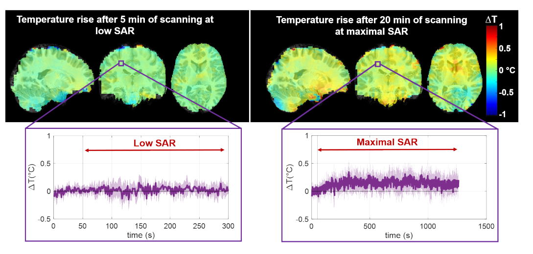
Measuring non-invasively the temperature rise occurring in cerebral MR exams.

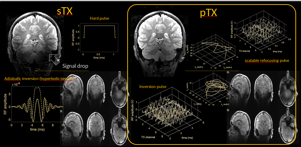
Parallel transmission technology

With 7 Tesla MRI (300 MHz), the image contrast (here we present 3D T2 weighted images with and without suppression of the fluid’s signal) is often impacted by the non-uniformity of the transmitted RF field. With a single transmit architecture (sTX, left image), this problem is difficult to mitigate, even with the use of high power adiabatic RF pulses for spin inversion. The parallel transmission technology (pTX), coupled with a pulse design strategy involving constrained non-convex (numerical) optimizations, can solve this problem while maintaining the RF energy deposition in the body low. The presented pTX pulses make use of parameterization-free RF and magnetic field gradient waveforms obtained by application of a GRAPE (Gradient Ascent Pulse Engineering) algorithm.

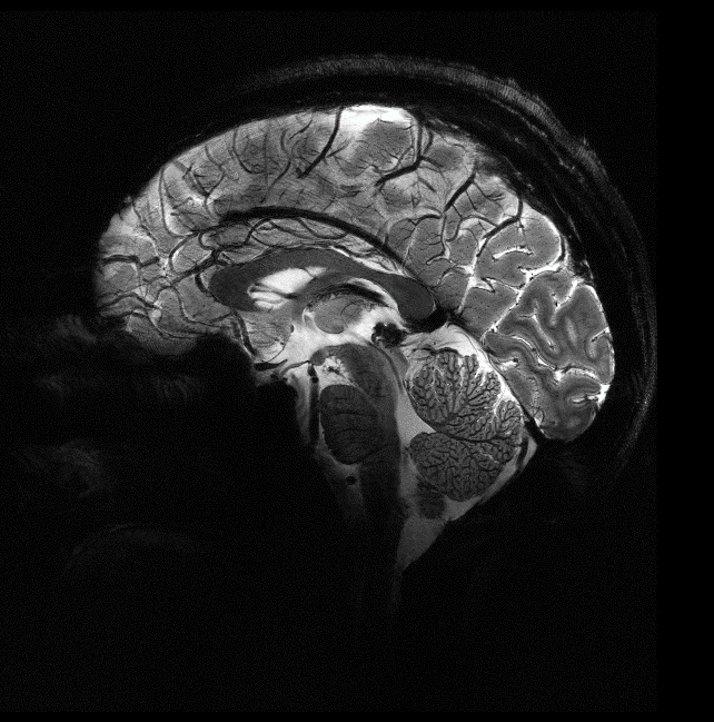
In vivo imaging at high resolution at 11.7T

Boulant, N., Mauconduit, F., Gras, V. et al. In vivo imaging of the human brain with the Iseult 11.7-T MRI scanner. Nat Methods 21, 2013–2016 (2024).