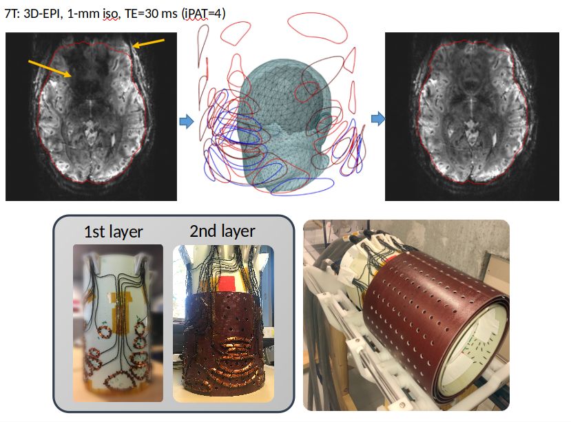
SCOTCH shim array
EPI imaging is improved thanks to the SCOTCH prototype - in this example, signal is recovered in the ventro-medial prefrontal cortex. Distortions along the phase-encode dimension are also reduced (EPI acceleration - CAIPIRINHA 2x2 in both images). Bottom left - first two layers of our 48-coil prototype. Bottom right - the three layers surround our home-made 8-Tx/32-Rx RF coil.
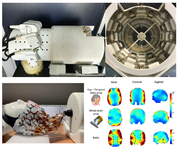
MRI at 11.7T
Top - The first in-vivo brain images at 11.7T were acquired with this 8-Tx/32-Rx Iseult coil composed of 15 dipoles, 16 loops and a patch. Bottom - An original honeycomb-shaped High Impedance Coil array was built on a neoprene cap to target Temporal Lobes fMRI at 11.7 T. Bottom right - SNR improvement over the temporal lobes thanks to this array.
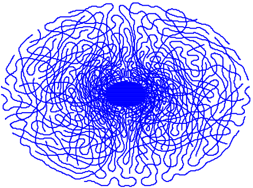
Sparkling
Iterative optimization of the Sparkling sampling for a 2D GRE. The final sampling fulfills Compressed Sensing criteria and is compatible with hardware limits (gradient amplitude and slew rate). Such an acquisition is highly accelerated compared to the fully sampled Cartesian acquisition (courtesy C. Lazarus, CEA BAOBAB & INRIA MIND)
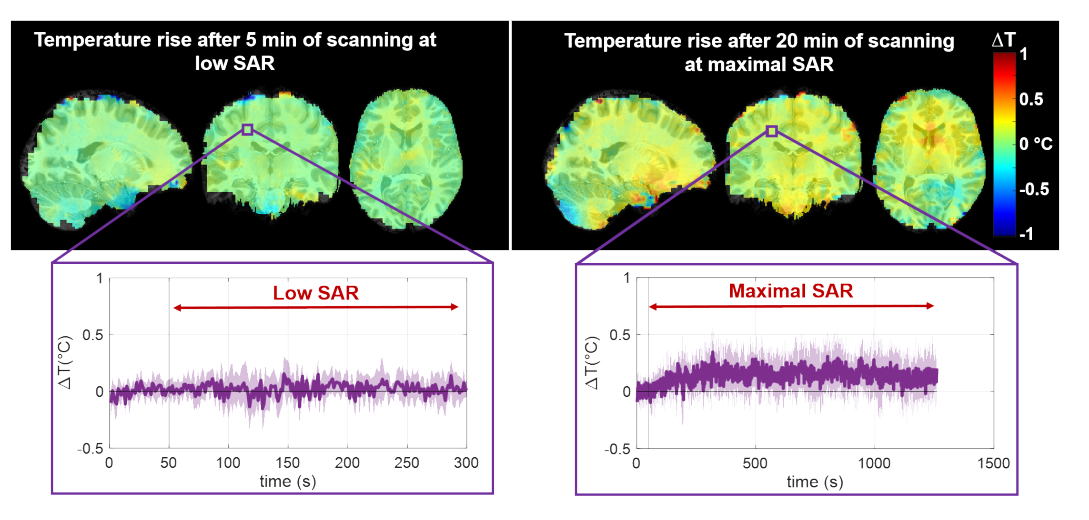
SAR
Measuring non-invasively the temperature rise occurring in cerebral MR exams.
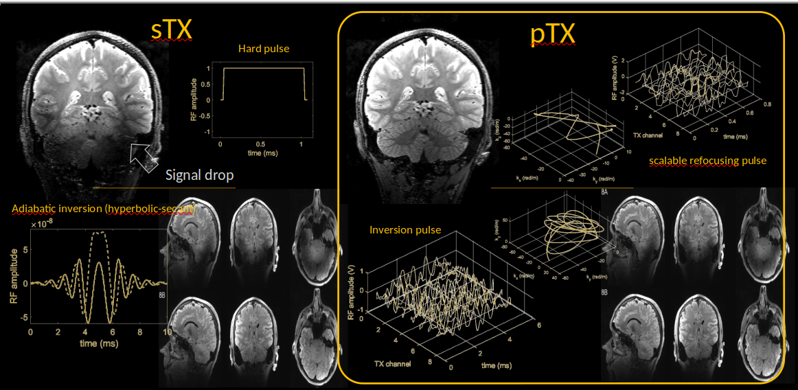
Parallel transmission technology
With 7 Tesla MRI (300 MHz), the image contrast (here we present 3D T2 weighted images with and without suppression of the fluid’s signal) is often impacted by the non-uniformity of the transmitted RF field. With a single transmit architecture (sTX, left image), this problem is difficult to mitigate, even with the use of high power adiabatic RF pulses for spin inversion. The parallel transmission technology (pTX), coupled with a pulse design strategy involving constrained non-convex (numerical) optimizations, can solve this problem while maintaining the RF energy deposition in the body low. The presented pTX pulses make use of parameterization-free RF and magnetic field gradient waveforms obtained by application of a GRAPE (Gradient Ascent Pulse Engineering) algorithm.
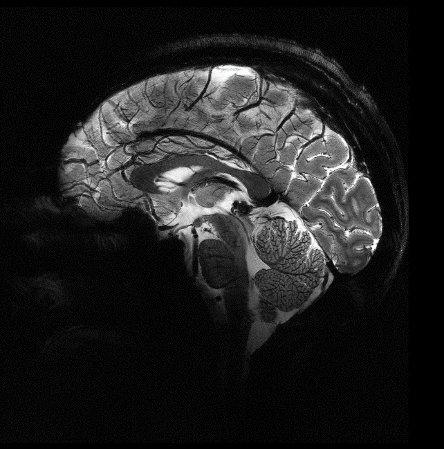
In vivo imaging at high resolution at 11.7T
Boulant, N., Mauconduit, F., Gras, V. et al. In vivo imaging of the human brain with the Iseult 11.7-T MRI scanner. Nat Methods 21, 2013–2016 (2024).